Retinal Disease Management

Managing & Treating Retinal Disease

Dr. Robert King and Dr. Robin Ray specialize in the evaluation and treatment of retinal diseases including macular degeneration, diabetic retinopathy, macular hole, macular pucker, and retinal detachment.

Retinal consultation is available in our Savannah, Bluffton, Brunswick, Glennville, Hinesville, Statesboro, Pooler and Vidalia locations.

Please call (912) 354-4800 for an appointment.

Common Conditions We Treat

Macular Degeneration

Cystoid Macular Edema

Diabetic Retinopathy

Retinal Vein/Artery Occlusion

Flashes + Floaters/Retinal Detachment

Epiretinal Membrane/Macular Pucker

The retina service utilizes the latest imaging modalities to diagnose and manage complex vitreoretinal pathology including:

Spectral-Domain Optical Coherence Tomography – Essential technology for the diagnosis and management of macular disease such as macular degeneration and diabetic macular edema).

Photo on the left is of a macula with edema. The photo on the right is a cross-sectional image (OCT) of the same macula showing the swelling (macular edema).

Photo on the left is of a macula with edema. The photo on the right is a cross-sectional image (OCT) of the same macula showing the swelling (macular edema).

  • Wide field imaging with the Optos California – Wide field imaging refers to new technology that allows us to take a single photo of your retina that routinely captures peripheral detail that can be missed with conventional retinal imaging. Standard photos are 30-to 50-degree views, but the new Optos system captures 200 degrees. The unique feature to the California model is that we can perform fluorescein and ICG angiography at the same time in evaluation of unusual inflammatory retinal conditions. It also increases the ability to discover peripheral vascular abnormalities that can be seen in diabetes and vein occlusion. This images are available in each exam room and are reviewed with the patient. This improved imaging has been very rewarding from a diagnostic standpoint but also in a patient’s understanding of his or her retinal problem. To be able to show a large, all-encompassing picture of the diabetic changes, retinal detachment, or tumor is invaluable in patient education.
  • Fluorescein Angiography – Imaging that uses dye injected into an arm in the vein to evaluate blood flow through the retina and identify areas of leakage in diseases such as diabetic retinopathy, macular degeneration, and vein occlusions.
angiog

Fluorescein angiography showing areas of poor blood flow (dark areas) in a patient with severe diabetic retinopathy.

  • Indocyanine Green Angiography – Imaging modalities that utilize special dye to evaluate the blood supply of the layer underneath the retina and pinpoint abnormal blood vessel formation in diseases such as macular degeneration and inflammatory conditions in the eye.
Right eye of a patient with Acute Posterior Multifocal Placoid Pigment Epitheliopathy (APMPPE), a rare inflammatory eye disease seen in young adults. The photo on the left is fluorescein angiography which highlights the blood vessels in the retina. The photo on the right is indocyanine green angiography which highlights the blood vessels under the retina.

Right eye of a patient with Acute Posterior Multifocal Placoid Pigment Epitheliopathy (APMPPE), a rare inflammatory eye disease seen in young adults. The photo on the left is fluorescein angiography which highlights the blood vessels in the retina. The photo on the right is indocyanine green angiography which highlights the blood vessels under the retina.

  • Fundus Autofluorescence – Useful in diagnosing and following diseases such as macular degeneration, retinitis pigmentosa, cone-rod dystrophies, plaquenil toxicity, Stargardt disease, etc.
Photo using special filters to show macular changes in a patient with retinitis pigmentosa.

Photo using special filters to show macular changes in a patient with retinitis pigmentosa.

  • High-Frequency Ultrasound – Essential for the evaluation of retinal anatomy in patients with dense cataracts or blood in the eye).
Ultrasound showing a retinal detachment in a patient with severe diabetic retinopathy.

Ultrasound showing a retinal detachment in a patient with severe diabetic retinopathy.

  • Ultrasound Biomicroscopy – Useful for evaluating tumors and complex anatomy that can predispose to glaucoma.
High-resolution ultrasound that shows details of the anterior segment (front part of the eye) in cross-section.

High-resolution ultrasound that shows details of the anterior segment (front part of the eye) in cross-section.

Flashers, Floaters and Retinal Detachment

floaters

What are flashes and floaters? What causes them?

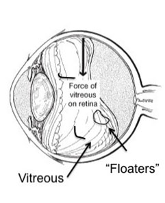
The center of the eye is filled with a clear gel called vitreous. When we are born, the vitreous in both eyes is completely clear and stuck down everywhere to the inside surface of the eye (including the retina). As we get older the gel clumps together – these cast shadows on the retina (light sensing part of the eye) causing the occasional “floaters” we see.

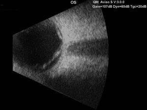
After several decades (usually after age 50) the gel liquefies and starts to separate from the back of the eye. This is called a posterior vitreous separation or detachment. When this occurs a large floater can be seen. People sometimes describe this as “a circle,” “a cobweb,” “a spider,” etc.

As the separation progresses (usually over 4-6 weeks) the gel can tug on the retina (back of the eye). This causes flashes of lights. Though the flashes do not cause damage, a flash could indicate that the gel is pulling especially hard increasing the risk of a tear in the retina.

The appearance of flashes and floaters may be alarming, especially if they develop suddenly. You should contact your eye doctor right away if you suddenly develop new flashes or floaters.

Are floaters ever serious?

The retina can tear if the shrinking vitreous gel pulls away from the wall of the eye. This sometimes causes a small amount of bleeding in the eye that may appear as new floaters. A torn retina is always a serious problem, since it can lead to a retinal detachment.

You should call your eye doctor as soon as possible if:

  • Even one new floater appears suddenly
  • Lots of “pepper spots” in your vision
  • You see sudden flashes of light
  • You notice loss of any peripheral (side) vision

How are your eyes examined for flashes and/or floaters?

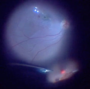
When an ophthalmologist examines your eyes, your pupils will be dilated with eye drops. During this examination, your ophthalmologist will carefully observe your retina and vitreous using a bright light. Evaluation for retinal tears usually requires a technique called “scleral depression” for which your ophthalmologist will use an instrument to gently press on your eyelids. This office examination is done with minimal discomfort.

Because your eyes have been dilated, you may need to make arrangements for someone to drive you home afterward. Floaters and flashes of light become more common as we grow older. While not all floaters and flashes are serious, you should always have a medical eye examination by an ophthalmologist to make sure there has been no damage to your retina.

What can be done about floaters?

Floaters can get in the way of clear vision, which may be quite annoying, especially if you are trying to read. Though most floaters will always be there, people usually notice them less over time. Floaters can be surgically removed, but this is not performed routinely as most people have minimal symptoms after a couple months. Even if you have had some floaters for years, you should have an eye examination immediately if you notice new ones.

What is a retinal detachment?

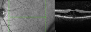
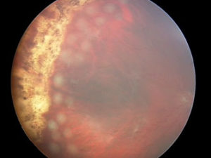
The retina is the sensory tissue that lines the back wall of the eye. It can be described as the wallpaper of the back of your eye. A retinal detachment occurs when the retina is separated from the inside of your eye and can no longer transmit images properly through the optic nerve to the brain. The center of the retina is the macula. If the macula is involved in the detachment, the vision is often extremely blurred. If the macula is not involved in the area of detachment, there may be some loss of peripheral vision.

Is retinal detachment serious?

Yes. A retinal detachment is a very serious problem that will almost always lead to blindness if not treated.

What causes a retinal detachment?

As described above, in some cases vitreous separation can cause a tear (or tears) in the retina. As time passes, fluid from the center of the eye will pass through the tear and begin accumulating beneath the retina, causing a retinal detachment. There are some conditions that increase the likelihood of developing a retinal detachment, including the following:

  • Myopia (nearsightedness)
  • Traumatic injury
  • A history of retinal detachment in either eye
  • Family history of retinal detachment

What are the symptoms of a retinal detachment?

  • A new floater or several floaters (can be hundreds) that appear suddenly
  • Sudden flashes of light in the eye
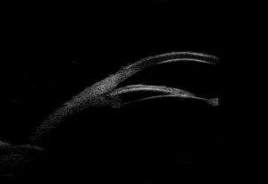
  • Seeing “cobwebs” or hairs in field of vision
  • Impression of a curtain or veil coming down over vision (Loss of peripheral, and then central,
    vision)
  • Blurred vision
  • Pain is not a symptom of a detached retina

How is retinal detachment repaired?

A retinal detachment can be repaired in several different ways. The surgery chosen depends on the age of the patient, the type of retinal detachment, whether the patient has had cataract surgery or not, surgeon preference, and other factors. The four general categories of retinal detachment repair are:

  • Laser barricade – does not fix the detachment but prevents it from spreading; can be done in the office).
Laser barricade

Laser barricade

  • Pneumatic Retinopexy – a combination of treating the tear with cryotherapy (freezing treatment) or laser and the use of a gas bubble injected into the eye to help the eye get rid of the fluid. The picture shows the injection of a gas bubble that will cover the tear in the retina. This prevents fluid from getting behind the retina and allows the body to absorb the fluid and flatten the retina.
Pneumatic Retinopexy

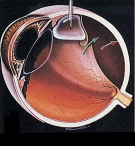
Pneumatic retinopexy

  • Scleral Buckle – a scleral buckle involves first treating the tear in the retina with cryotherapy (freezing treatment) and then placing a silicone band under the muscles around the eye to support the tear. The fluid can be drained from under the retina or a gas bubble can be injected into the eye similar to pneumatic retinopexy.
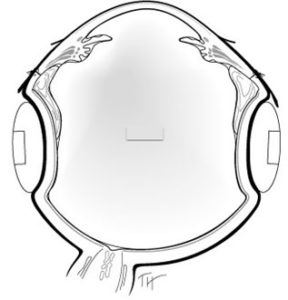
Scleral Buckle

Scleral Buckle

  • Vitrectomy – vitrectomy involves the removal of the vitreous (floaters) from the eye using small instruments. This removes all the traction (pulling) on the retinal tear(s) and detachment. The vitreous space is then filled with a long-lasting gas bubble that keeps the retina flat until the tears heal. Laser use used to seal the tear(s).
Vitrectomy

Vitrectomy

Sometimes both a scleral buckle procedure and vitrectomy are used together to fix complex retinal detachments.

For additional information, download the following forms about:

  • Diabetic Retinopathy
  • Epiretinal Membrane/Macular Pucker
  • Flashes, Floaters and Retinal Detachment

Macular Pucker

What is an epiretinal membrane?

An epiretinal membrane is an abnormal layer of tissue found on the surface of the retina which is the inside surface of the back of the eye. Specifically, this forms on the “macula” which is the center of the retina. This layer can “contract” and pull on the surface causing distortion to the normal structure of the macula. This pulling can cause swelling which leads to irreparable damage to vision.

Normal Macula
Epiretinal Membrane causing distortion of the Macula

What types of symptoms do patients with epiretinal membranes have?

Symptoms vary based on the severity of the swelling/traction caused by the membrane. The most typical symptoms are:

  • Distortion of vision (called “metamorphopsia”) – letters look crooked
  • Blurred vision or loss of visual acuity
  • Dark or “gray” spots near center of vision – patients describe missing letters in words when looking right at the word. These dark spots are different than “floaters” as they do not move around.

What causes an epiretinal membrane?

There are several causes of epiretinal membrane. The most common reason is a posterior vitreous separation or detachment. This is when the vitreous gel that fills the center of the eye separates from the back surface of the inside of the eye (the retina). A layer of gel can remain behind and surface as “scaffold” for cells to grow and then contract. This contraction causes the surface to become irregular and for the vision to worsen.

Other causes of epiretinal membrane are:

  • Retinal tear/detachment (or after surgery for these conditions)
  • Inflammation in the eye (“Uveitis”)
  • Bleeding in the eye (from diabetes, retinal tears, abnormal blood vessel growth from
    other conditions)

How are epiretinal membranes diagnosed?

The macula can be visualized during a routine eye exam after dilation of the pupil. Optical coherence tomography is a type of non-invasive imaging that allows your physician to view the macula and epiretinal membrane (if present) in cross-section. This allows your physician to see how much swelling and distortion is present. This is useful for following the disease over time.

Undilated Pupil
Dilated Pupil
View of retina/macula (back of eye) through Dilated Pupil
Cross sectional image (Optical Coherence Tomography, or OCT) of a normal macula. The “dip” in the center is the very center part of the vision, or the “fovea.” This is the “20/20? portion of the vision.
Cross sectional image (OCT) of a macula with an epiretinal membrane. Notice the loss of the “dip” and the distortion to the retinal surface. The retina is also thicker because of the membrane. This distortion and thickening leads to decreased vision.

My eye doctor told me I have a macular pucker. How is that different?

There are several different terms that essentially mean the same thing:

  • Epiretinal Membrane (ERM)
  • Epimacular Membrane
  • Macular Pucker
  • Cellophane Retinopathy
  • Vitreomacular Traction (VMT) – implies the vitreous gel is pulling the retina towards the front of the eye
  • Vitreomacular Adhesion Syndrome (same as VMT)

How common are epiretinal membranes?

Epiretinal membranes are quite common but they do not always cause changes in vision. Many times an epiretinal membrane is an incidental finding on a routine eye exam. Patients with normal vision and minimal changes to the retina/macula can be observed safely.

How are epiretinal membranes treated?

If visually significant, epiretinal membranes are treated with surgery. No oral medications or eye drops will help this condition. Surgery consists of “vitrectomy” to remove the vitreous gel (“floaters”) from the middle of the eye. Micro-forceps are then used to gently peel the membrane away from the retinal surface.

Removal of the vitreous from inside the eye using the “vitrector” (left side of picture). A small instrument (right side of picture) provides the light to see inside the eye. This is done under a microscope.
Gently peeling of the epiretinal membrane from off the retinal surface using micro-forceps. This relieves the traction causing the distortion and swelling of the retina/macula. What is the vision like after surgery? The vision is generally quite blurry the day after surgery. Patients normally notice a return of their pre-operative vision 2-4 weeks after surgery. An improvement in the distortion is usually the first thing patients notice. The best possible vision may not come for 9-12 months after surgery.

What are the risks of surgery?

Any intraocular surgery involving vitrectomy has a low risk of infection, bleeding, retinal detachment, and other complications, which can all lead to permanent vision loss. Risks, benefits, and alternatives to surgery should be discussed with your surgeon prior to proceeding with vitrectomy for epiretinal membrane.