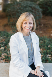
Georgia Eye Institute Welcomes Dr. Alison Pott
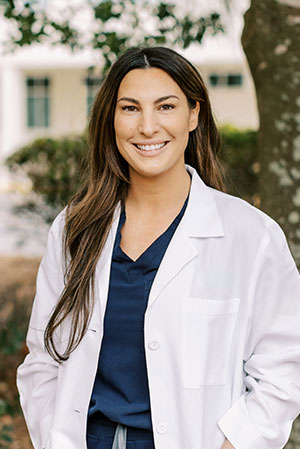
February 11th, 2026 – Savannah, GA – Georgia Eye Institute (GEI) announces the arrival of Dr. Alison Pott to the practice. She is an optometrist with a focus on comprehensive eye care, including disease management services.
Dr. Pott earned a Bachelors of Science in Intergrative Physiology at the University of Colorado, Boulder before completing her Doctor of Optometry at Midwestern University in Arizona. She has experience working with patients of all ages, including managing myopia progression, strabismus amblyopia, and keratoconus in children as well as diabetic retinopathy and glaucoma management in adults. She is now accepting patients at the Savannah location.
Georgia Eye Institute provides primary care, optical retail locations, and sub-specialty eye care in 10 locations throughout Southeast Georgia and South Carolina. Subspecialty eye care includes cataract evaluations, retinal care, glaucoma management, functional cosmetic eye surgery, and LASIK laser vision correction.
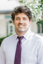
Dr. Brandon Fram Joins Georgia Eye Institute
October 6th, 2025 – Savannah, GA – Georgia Eye Institute (GEI) announces the arrival of Dr. Brandon Fram to the practice. He is an Ophthalmologist specializing in retinal diseases and surgery.
Dr. Fram received his undergraduate degree from the University of Georgia before attending Mercer University School of Medicine for his doctorate. From there, he completed his residency in Ophthalmology at Virginia Commonwealth University Health System in Richmond, Virginia, where he served as Chief Resident, and his surgical Retina Fellowship at Retina and Vitreous of Texas, in Houston. His research has been published in multiple medical journals, including the Journal of Vitreoretinal Diseases, the American Journal of Ophthalmology, and Retina Today. Dr. Fram is now accepting patients in Savannah, Pooler, Hinesville, and Statesboro.
Georgia Eye Institute provides primary care, optical retail locations, and sub-specialty eye care in 11 locations located throughout Southeast Georgia and South Carolina. Subspecialty eye care includes cataract evaluations, retinal care, glaucoma management, functional cosmetic eye surgery, and LASIK laser vision correction.
Dr. Christopher Richmond Joins Georgia Eye Institute
June 28, 2023 – Savannah, GA – Georgia Eye Institute (GEI) announces the arrival of Dr. Christopher Richmond to the practice. Dr. Richmond is an Ophthalmologist specializing in cataract and refractive surgery.
Dr. Richmond received his undergraduate degree from Clemson University before attending the University of South Carolina School of Medicine for his doctorate. He completed his internship in General Surgery at Walter Reed National Military Medical Center and his residency in Ophthalmology at the Naval Medical Center San Diego. Dr. Richmond is a member of the American Academy of Ophthalmology and the Society of Military Refractive Surgeons.
Prior to joining Georgia Eye Institute, Dr. Richmond served as an officer and a flight surgeon in the United States Navy. Most recently, he was the Department Head of Ophthalmology at the Naval Hospital in Pensacola, Florida. Dr. Richmond is currently accepting patients at the Savannah campus.
Georgia Eye Institute provides primary care, optical retail locations, and sub-specialty eye care in 11 locations located throughout Southeast Georgia and South Carolina. Subspecialty eye care includes cataract evaluations, retinal care, glaucoma management, functional cosmetic eye surgery, and LASIK laser vision correction.
Dr. Nicolle Oberlin Joins Georgia Eye Institute
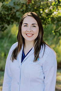
April 5, 2022 – Savannah, GA – Georgia Eye Institute (GEI) announces the arrival of Nicolle Oberlin to the staff. Oberlin is an Optometrist with special interests in primary care, including pediatrics and disease, as well as military optometry.
Oberlin received her Bachelor of Science-Vision Science degree and her Doctor of Optometry degree from the College of Optometry at Ferris State University, in Big Rapids, Michigan. She is an active member of the Armed Forces Optometric Association, Michigan Optometric Association, and the American Optometric Association.
Oberlin has previously done an externship at a Special Needs Vision Clinic in Michigan, and most recently was active-duty Assistant Chief of Optometry at Winn Army Community Hospital in Fort Stewart, Georgia. She is now accepting patients in the Hinesville location.
Georgia Eye Institute provides primary care, optical retail locations, and sub-specialty eye care in 12 locations located throughout Southeast Georgia and South Carolina. Our subspecialty eye care includes cataract evaluations, retinal care, glaucoma management, functional cosmetic eye surgery, and LASIK laser vision correction.
Dr. William Degenhart Receives Beacon of Hope Award from Georgia Lions Lighthouse
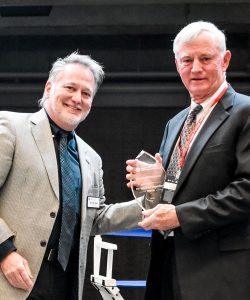
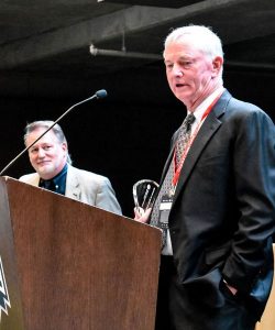
October 19, 2018 – Savannah, GA – Dr. William Degenhart, board-certified ophthalmologist and founding partner of Georgia Eye Institute, received the Georgia Lions Lighthouse Beacon of Hope Award last night at their annual Evening in the End Zone event held in Atlanta.
The Georgia Lions Lighthouse Beacon of Hope Award honors a medical professional who demonstrates an unwavering commitment to vision or hearing care for the uninsured in his or her community. Dr. Degenhart has been providing eye care since 1982 and his volunteer efforts and support of those in need throughout Savannah and Southeast Georgia continue to inspire and encourage many.
Georgia Eye Institute provides primary eye care, optical retail locations and sub-specialty eye care in 13 locations conveniently located throughout Southeast Georgia and the South Carolina. The sub-specialty eye care includes cataract evaluation and management, retinal care, glaucoma management, functional cosmetic eye surgery and LASIK laser vision correction.
Dr. Thomas James Kandl Joins Georgia Eye Institute
September 6, 2018 – Savannah, GA – Georgia Eye Institute (GEI) announces the addition of Dr. Thomas James Kandl to their staff. Kandl has completed a fellowship in Ophthalmic Plastic and Reconstructive Surgery at the MD Anderson Cancer Center in Houston, Texas. He is seeing patients at the main office in Savannah and specializes in Oculoplastics and Reconstructive Surgery.
Kandl received a Bachelor of Science Degree from Wofford College; a Doctor of Medicine Degree from the Medical University of South Carolina; and was awarded a Doctor of Ophthalmology Degree from Rutgers University.
Georgia Eye Institute provides primary eye care, optical retail locations and sub-specialty eye care in 13 locations conveniently located throughout Southeast Georgia and the South Carolina. The sub-specialty eye care includes cataract evaluation and management, retinal care, glaucoma management, functional cosmetic eye surgery and LASIK laser vision correction.
Georgia Eye Institute is Working To Promote Eye Exams in Southeast Georgia and the Low Country
May 2018 – Millions of people in the United States have undetected vision problems that can cause vision loss and even blindness. Unfortunately, many eye diseases have no early warnings signs or symptoms so you may be affected without even noticing it. Visiting your eye care professional for a comprehensive dilated eye exam is the only way to know if your vision is at its best and your eyes are healthy.
A comprehensive dilated eye exam is a painless procedure in which drops are placed in your eyes to dilate, or widen, the pupil. This allows your eye care professional get a good look at the back of your eyes and examine them for any signs of damage or disease. Georgia Eye Institute recommends that you put a visit to your eye care professional on your “to do” list. Detecting eye diseases in their early stages can help save your sight.
Comprehensive dilated eye exams are important for maintaining good eye health. Healthy vision can help keep you safe when you are behind the wheel, while you are participating in sports, or during recreational activities. It can also help you perform your best on the job and ensure that you maintain a healthy and active lifestyle well into your golden years.
Take care of your eyes and they will help take care of you. Find a window of time to schedule an eye exam today.
Dr. Shari Carney Joins Georgia Eye Institute
28 February 2017 – Savannah, GA – Georgia Eye Institute (GEI) announces the addition of Dr. Shari A. Carney to their staff. Carney, a medical ophthalmologist, will begin seeing patients at the main campus of GEI on February 19th.
Carney received a Bachelor of Science Degree from Clarkson University and a Doctorate of Medicine from Upstate Medical University where she was later Chief Resident in Ophthalmology. Most recently, she was Clinical Assistant Professor of Medical Education for Mercer University School of Medicine, at Memorial University Medical Center. She is a member of the American Academy of Ophthalmology.
Georgia Eye Institute provides primary eye care, optical retail locations and sub-specialty eye care in 13 locations conveniently located throughout Southeast Georgia and the South Carolina. The sub-specialty eye care includes cataract evaluation and management, retinal care, glaucoma management, functional cosmetic eye surgery and LASIK laser vision correction. For more information, visit gaeyeinstitute.com.
Eye Safety for the Solar Eclipse

By Mark Manocha, M.D.
During August 21st’s solar eclipse, it is important to take the proper ocular safety precautions. Looking directly at the sun is never a good idea, so there are a few ways to view it and minimize risk. Savannah will only experience a partial solar eclipse (around 90%), but you can look online to see where totality occurs.
- At no point in Savannah will it be safe to look directly at the sun because it will never be fully covered. Even when the sun is partially covered, it is just as bright, and is equally unsafe.
- Put on your glasses immediately after the sun emerges as looking at it directly, however brief, can cause long-term retinal damage.
- Make sure to look away from the sun before putting on and removing your protective glasses; rapid adjustment to light can also cause severe eye damage.
- You can purchase specially certified glasses (ISO 12312-2) for viewing, which you can find online or in certain stores.
- Regular sunglasses, cameras, binoculars, and telescopes are not safe for viewing the eclipse.
- Alternatively, you can use the pinhole method to project an image of the eclipse on another material. You can do this by poking a hole in a piece of cardboard, and putting a piece of paper behind it, changing the distance until the image is projected clearly onto it.
The solar eclipse is one of the greatest events nature can offer, and it happens rarely in United States. Don’t let this opportunity to witness history pass you by, but make sure to be safe and protect your eyes while viewing this grand spectacle.
Basics of Glaucoma
More than 3 million people in the U.S. have glaucoma. Glaucoma is a complicated eye disease in which the optic nerve gets damaged due to high eye pressure. If it’s not treated properly or is ignored glaucoma can lead to progressive, irreversible loss of vision. Glaucoma is the second most common cause of blindness in the world today.
Glaucoma is incurable, but it can be managed through drug therapies. However, when medication fails to lower the eye pressure, or causes severe side effects, surgery is recommended. In some cases, doctors might recommend surgery as the first course of treatment.
Glaucoma surgery is typically categorized two ways: laser surgery; and incisional surgery. Your ophthalmologist will take into consideration the type of glaucoma you have, its severity, and your general eye health before deciding which type of surgery is most suitable for your condition.
Surgery can stabilize vision by lowering the eye pressure, but it cannot cure glaucoma or reverse any loss of vision which has already occurred.
The following is a brief overview of glaucoma laser surgery and incisional surgery, and what to expect during recovery.
Laser Surgery
Doctors ordinarily recommend laser surgery unless pressure in the eye is extremely high or the damage to the optic nerve is severe.
During this procedure, the eye surgeon improves the drainage system of the eye using a highly focused beam of light. The patient feels no pain during the surgery, as the eye is numbed. A special lens is held to the eye and the laser beam is directed into it.
Immediately after the surgery you may experience blurry vision and some irritation of the eye. Many patients are able to resume normal activities within a day or two after the surgery. However, most doctors recommend patients not lift anything heavy, refrain for any strenuous activity, or do any significant bending for at least two weeks.
Incisional Surgery
Your eye doctor may recommend incisional surgery for any of the following three conditions:
- Extremely high eye pressure
- A severely damaged optic nerve
- Failure to lower eye pressure with laser surgery
During incisional surgery, a small drainage hole is made in the sclera to allow intraocular fluid to bypass the clogged drainage canals and allow the fluid flow from the eye, helping reduce eye pressure.
Shortly after incisional surgery, patients may experience irritation, redness, and increased watering or tearing. Recovery time for incisional surgery is typically longer as compared to laser surgery. Most patients recover in two to four weeks. In some cases, however, recovery time might be longer – up to 2 months.
For more information please call 912.354.4800.